

視野検査
視神経の検査 (perimetry:英)
視野検査(しやけんさ)とは、「まっすぐ一点を見つめた状態で、上下左右どのくらいの広い範囲が見えているか」、また「それぞれの場所でどの程度の明るさまで認識できるか(網膜の感度)」を測定する検査です。
視力が「形をはっきりと見分ける能力」であるのに対し、視野は「見える空間の広がり」を指します。片方の目で見えない部分があっても、もう一方の目が補ってしまうため、視野の欠けは自分では気づきにくいのが特徴です。
A visual field test (perimetry) is an examination to measure the extent of the area you can see (up, down, left, and right) while keeping your gaze fixed on a single point straight ahead. It also measures the level of brightness you can perceive at each location (retinal sensitivity).
While visual acuity is the "ability to clearly distinguish shapes," the visual field refers to the "extent of the visible space." A key characteristic of the visual field is that even if there is a missing area in the vision of one eye, it is difficult to notice on your own because the other eye compensates for it.
視野検査でわかる疾患と見え方の特徴
What a Visual Field Test Reveals
視野の欠け方(欠損)や狭まり方のパターンを分析することで、目から脳に至る経路のどこに異常があるかを特定できます。
By analyzing the patterns of vision loss (defects) or narrowing, we can identify where abnormalities exist along the pathway from the eyes to the brain.
By analyzing the patterns of vision loss (defects) or narrowing, we can identify where abnormalities exist along the pathway from the eyes to the brain.
| 対象部位 Target Area |
主な疾患例 Example of Condition |
視野(見え方)の変化 Changes in Vision |
| 目(全体) Eyes (General) |
緑内障 Glaucoma |
初期は無症状だが、少しずつ視野の一部が欠け始める。最重要の検査対象。 No symptoms in the early stages, but small parts of the visual field gradually begin to disappear. |
| 視神経 Optic Nerve |
視神経炎など Optic Neuritis, etc. |
視野の中心部が見えにくくなる(中心暗転)。 Difficulty seeing in the center of the visual field (central scotoma). |
| 網膜 Retina |
網膜剥離・網膜色素変性症 Retinal Detachment, Retinitis Pigmentosa |
視野が狭くなったり、部分的に欠けたりする。 The visual field becomes narrowed or partially missing. |
| 脳 Brain |
脳梗塞・脳腫瘍 Cerebral Infarction (Stroke), Brain Tumor |
左右どちらかの半分が見えなくなる「半盲(はんもう)」が起こる。 "Hemianopsia" may occur, where one half (left or right) of the visual field is lost. |
主な検査の種類
Main Types of Visual Field Tests
目的に応じて、主に以下の2種類の装置が使われます。
Depending on the purpose, the following two types of devices are mainly used.
Depending on the purpose, the following two types of devices are mainly used.
| 検査の種類 Types of Test |
主な装置名 Common Device Names |
特徴・測定方法 Features & Methodology |
| 静的視野検査 Static Perimetry |
ハンフリー等 Humphrey, etc. |
一定の範囲内に、強さ(明るさ)の異なる光を提示して感度を測る。 Measures sensitivity by presenting lights of varying intensity (brightness) within a fixed range. |
| 動的視野検査 Kinetic Perimetry |
ゴールドマン等 Goldmann, etc. |
動く光を周辺から中心へ移動させ、見える範囲の「等高線」を描く。 Draws "contour lines" of the visible range by moving a moving light from the periphery toward the center. |
検査の流れとポイント
Procedure and Important Notes
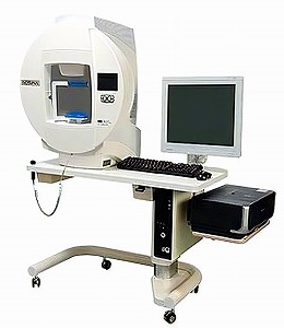
検査は暗い部屋で行われ、片目ずつ測定します。
The test is performed in a dark room, and each eye is measured separately.
The test is performed in a dark room, and each eye is measured separately.
| 検査のステップ Steps of the Test |
||
| 1 | 固定 Positioning |
顔を器械に固定し、中央にある固定された点を見つめる。 Your face will be secured to the machine. Keep your gaze fixed on the central target point. |
| 2 | 反応 Reaction |
視線は動かさず、周辺に光が見えた瞬間に手元のボタンを押す。 Without moving your eyes, press the button in your hand the instant you see a light in your peripheral vision. |
| 3 | 時間 Duration |
片目につき5分〜15分程度。 The test takes approximately 5 to 15 minutes per eye. |
受診時のアドバイス
Tips for the Patient
・迷わず押す:「見えた!」と思ったら迷わず押すのがコツです。
・休憩OK:集中力が必要な検査ですが、疲れたら合図をして休憩することも可能です。
・瞬きは自然に:瞬きは我慢せず、自然にしても大丈夫です(器械が調整してくれます)。
・Don't Hesitate: The key is to press the button without hesitation as soon as you think, "I saw it!"
・Take Breaks: This test requires concentration, but you can signal the staff to take a break if you feel tired.
・Blink Naturally: It is okay to blink normally; the machine is designed to adjust for it.