

OCT検査
OCT検査 (Optical Coherence Tomography scan:英)
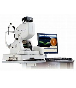
OCT(光干渉断層計)検査とは、赤外線を利用して目の奥(網膜)の断面図を撮影する検査のことです。
簡単に言うと「目のCTスキャン」のようなもので、表面からでは見えない網膜の内部構造や、視神経の厚みをミリ単位(ミクロンレベル)の精度で詳しく調べることができます。
An OCT (Optical Coherence Tomography) scan is a diagnostic test that uses infrared light to capture cross-sectional images of the back of the eye (the retina).
Put simply, it is like a "CT scan for the eye." It allows for a detailed examination of the internal structure of the retina and the thickness of the optic nerve—areas not visible from the surface—with precision down to the millimeter (micron) level.
1. OCT検査でわかること(対象疾患)
1. What can be detected with OCT?
従来の眼底カメラが「表面」を撮るのに対し、OCTは「断層(厚みや重なり)」を撮るため、以下の早期発見に有効です。
While conventional fundus cameras take "surface photos," OCT captures "cross-sectional layers." This makes it highly effective for the early detection of the following conditions:
While conventional fundus cameras take "surface photos," OCT captures "cross-sectional layers." This makes it highly effective for the early detection of the following conditions:
| 対象疾患 Condition |
検査でチェックするポイント What we check |
| 緑内障 Glaucoma |
視神経の厚みを測定。視野が欠ける前の、ごく初期段階で発見可能。 We check for thinning of the optic nerve. Detection is possible at the very early stage, even before vision loss begins. |
| 加齢性黄斑変性 Age-related Macular Degeneration |
網膜の中心(黄斑)に異常な血管やむくみがないかを確認。 We check for abnormal blood vessels or swelling in the center of the retina (the macula). |
| 糖尿病網膜症 Diabetic Retinopathy |
網膜のむくみ(浮腫)や血管の状態を詳しく確認。 We confirm the presence of swelling (edema) in the retina and assess the condition of the blood vessels. |
| 黄斑円孔・網膜剥離 Macular Hole/Retinal Dtachment |
網膜に穴が開いたり、剥がれたりしていないかを立体的に確認。 We can provide a 3D confirmation of whether there are any holes or peeling in the retina. |
2. OCT検査の3つのメリット
Key Features of the OCT Scan
患者さんの負担が少ないのがこの検査の大きな特徴です。
The scan is designed to be quick and stress-free for the patient.
The scan is designed to be quick and stress-free for the patient.
| 項目 Feature |
内容 Description |
| 痛みがゼロ Painless |
目に機械が触れることはありません。台にあごを乗せて中の光を数秒間見るだけです。 The device dose not touch your eye. Simply rest your chin on the stand and look at a light for a few seconds. |
| スピーディー Quick |
撮影自体は数秒。準備を含めても5〜10分程度で終わります。 The entire process takes about 5-10 minutes including preparation. The scan itself is finished in seconds. |
| 眩しくない Less Glare |
赤外線を使うため、通常の眼底検査に比べて眩しさを感じにくい. Because it uses infrared light, it is much less dazzling than a standard fundus examination. |